
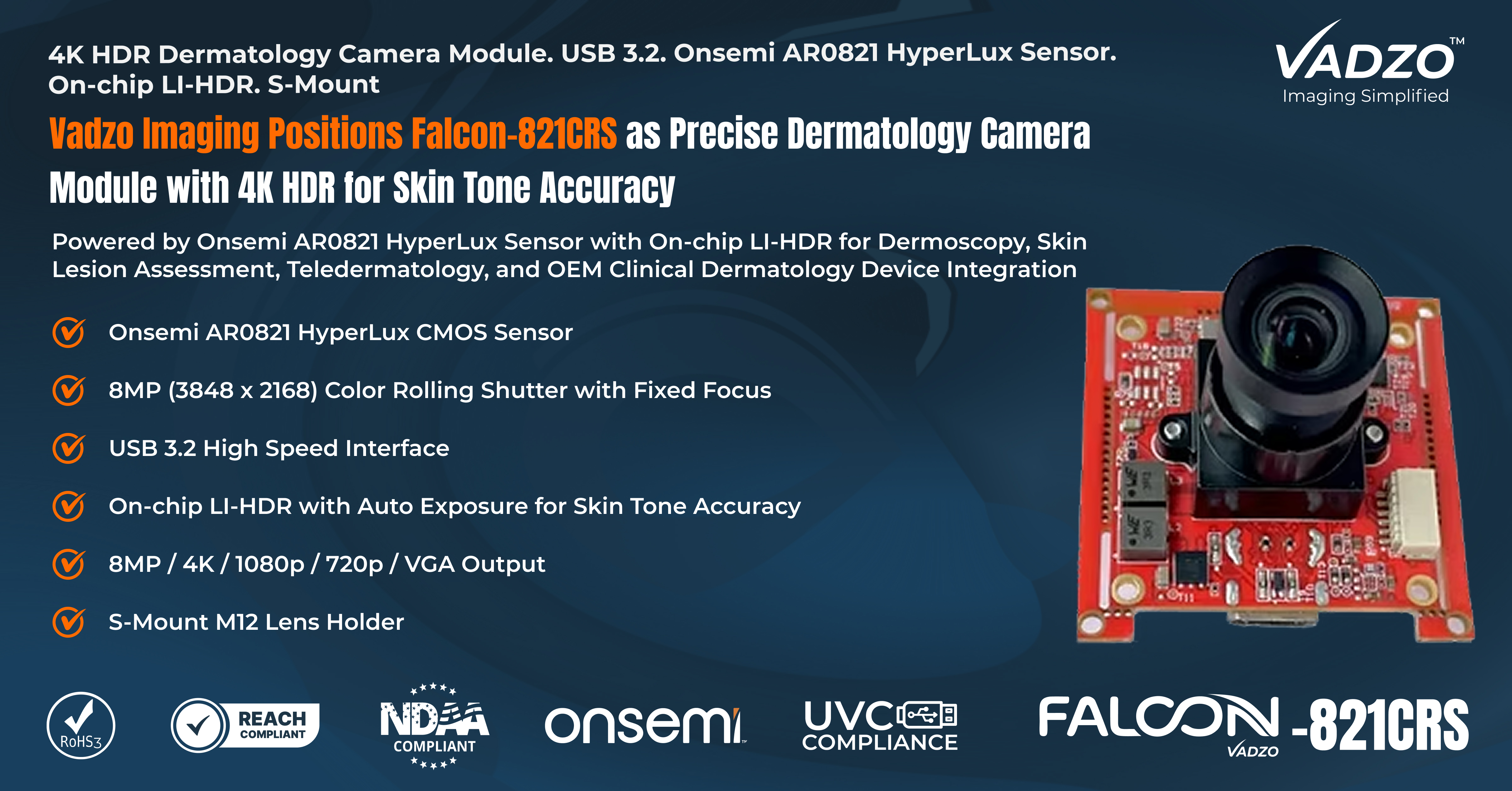
Vadzo Imaging Positions Falcon-821CRS as Precise Dermatology Camera Module with 4K HDR for Skin Tone Accuracy
The Falcon-821CRS is an 8MP 4K HDR dermatology camera module built on the Onsemi AR0821 HyperLux sensor. Positioned for dermoscopy, skin lesion assessment, teledermatology platforms, and clinical dermatology workflows, this color-accurate USB camera module delivers 4K output with on-chip High Dynamic Range, precise color reproduction, and USB 3.2 connectivity. It supports Windows, Linux, and Android in an S-Mount form factor, offering OEM dermatology device designers and clinical imaging engineers a production-ready platform built specifically for skin tone accuracy.
FORT WORTH, TX / ACCESS Newswire / June 29, 2026 /Vadzo Imaging, a provider of embedded vision camera products, today positions the Falcon-821CRS as a precision Dermatology Camera Module. Built on the Onsemi AR0821 HyperLux sensor and part of Vadzo 's medical imaging portfolio, this AR0821 Dermatology Camera delivers 8MP 4K color imaging with on-chip LI-HDR and USB 3.2 connectivity, targeted at dermoscopy systems, teledermatology platforms, and point-of-care skin imaging equipment. The Falcon-821CRS addresses a fundamental challenge in dermatology imaging: skin tone accuracy demands that the camera reproduce the visible color spectrum with high fidelity across the full range of human skin phototypes. A camera that introduces color bias or compresses dynamic range incorrectly changes the apparent appearance of lesions and skin conditions, undermining the clinical value of the captured image. This AR0821 Skin Imaging Camera combines the AR0821 sensor 's accurate color pipeline with on-chip LI-HDR and USB 3.2 integration in a single compact module. As an AR0821 Dermatology USB Camera, the Falcon-821CRS delivers the color precision and spatial resolution that dermatology imaging workflows require, from clinical consultation to teledermatology review.
Sensor and Camera Overview
The Falcon-821CRS is a Dermatology Camera Module built on the Onsemi AR0821 HyperLux sensor and coupled with a high-performance ISP. The AR0821 is an 8MP (3840 x 2160) color rolling shutter CMOS sensor with a 1/1.7-inch optical format and 2.1 µm pixel size. The sensor 's color pipeline delivers accurate spectral response across the visible range, with fidelity in the red and near infrared bands that are diagnostically relevant for melanin-based lesions and vascular structures in skin tissue. The Onsemi AR0821 Dermatology Camera design captures color data with the accuracy that dermatology diagnostic workflows require, where a shift of several kelvin in color temperature or a deviation in green channel response can alter the perceived character of a pigmented lesion.
The Falcon-821CRS functions as an AR0821 Lesion Imaging Camera through its combination of the AR0821 sensor, a high-performance ISP, and an S-Mount (M12) lens holder in a compact form factor suited to dermatology device enclosure integration. The on-chip LI-HDR mode preserves detail across the full dynamic range of skin surfaces, from specular reflections on oily or wet skin under dermoscopy contact fluid to absorption in darkly pigmented lesions. This capability makes the Falcon-821CRS relevant for both standard color imaging and HDR Skin Imaging Camera applications in clinical dermatology. UVC class compliance means the camera enumerates on any USB 3.2 host running Windows, Linux, or Android without additional drivers, simplifying integration into dermatology workstations and tablet-based clinical systems. The Dermatology Imaging Module supports output modes that cover full 8MP, 4K, 1080p, 720p, and VGA, allowing system designers to match frame rate and resolution to the downstream display or documentation workflow.
Key specs: 8MP (3848 x 2168) | Onsemi AR0821 1/1.7 inch 2.1 µm pixel | Color | Rolling Shutter | Fixed Focus | on-chip LI-HDR | Auto Exposure | High-Performance ISP | USB 3.2 | 8MP / 4K / 1080p / 720p / VGA | S-Mount (M12) | Windows Linux Android | UVC Compliant
Key Capabilities of the Onsemi AR0821 4K HDR Dermatology Camera Module
Color Accurate Imaging for Dermatology Diagnostics:Dermatology diagnosis depends on the accurate visual rendering of skin color, pigment distribution, and vascular patterns within a lesion. Cameras with poor color pipelines introduce systematic bias in the red, green, and blue channel response, causing skin tones to shift in ways that alter the apparent appearance of melanocytic lesions. The AR0821 sensor 's color pipeline delivers an accurate spectral response that translates to faithful skin tone reproduction across Fitzpatrick phototypes through VI. This color-accurate USB Camera renders the subtle color gradients that differentiate benign nevi from early-stage melanoma and identifies erythematous patterns in inflammatory dermatoses. As a Skin Tone USB Camera, the Falcon-821CRS provides the color fidelity that allows clinicians to compare images over time with confidence that any color change observed in the documentation reflects actual tissue change rather than camera calibration drift. The consistent Color Reproduction USB Camera performance reduces the interpretation burden for remote dermatologists reviewing teledermatology images, where color accuracy is the primary proxy for the information they would obtain in a direct physical examination.
On-chip HDR for Skin Lesion Contrast and Detail:Skin surfaces present a wide dynamic range of challenges. Specular highlights on the skin surface, particularly on oily or wet skin under dermoscopy, coexist with darkly absorbing pigmented lesions within the same frame. Standard cameras clip the highlight regions and lose the surface texture context that surrounds lesion borders. The AR0821 's on-chip LI-HDR captures this full contrast range in a single exposure, preserving both the surface detail in bright areas and the pigment depth in dark lesion regions. This WDR Dermatology Camera approach delivers images where dermoscopy patterns, including pigment networks, regression structures, and vascular morphology, remain visible throughout the lesion area without highlight clipping disrupting border analysis. The result is a High Fidelity Color Camera output that provides clinicians with more complete lesion information per image than standard dynamic range capture methods can deliver, supporting more accurate lesion assessment under variable clinical lighting conditions.
8MP Spatial Resolution for Lesion Morphology Analysis:Dermoscopy and skin lesion assessment depend on resolving fine morphological structures: pigment network geometry, follicular patterns, regression of white areas, and vascular loop structures. These features range in size from approximately 0.1 mm to several millimeters within a single lesion. The Onsemi AR0821 delivers 8MP (3840 x 2160) spatial resolution with a 2.1 µm pixel pitch, providing the sampling density needed to capture these fine structures when paired with appropriate dermoscopy optics. This 4K Dermatology USB Camera preserves lesion morphology at the pixel level, enabling post-capture digital magnification without loss of clinically relevant structural detail. The 8MP Skin Imaging Camera output integrates directly with AI-assisted dermatology analysis tools that require full resolution input to train and apply pigment network classifiers and pattern recognition algorithms. For OEM designers building dermoscopy systems, the 8MP frame provides the resolution headroom necessary to support both diagnostic visualization and downstream computational analysis within the same image capture.
USB 3.2 Plug and Play Integration with Dermatology Workstations:Dermatology clinics and point-of-care environments operate a wide range of host platforms, from general-purpose Windows workstations to dedicated dermatology imaging software running on Android laptops. Custom drivers introduce IT management overhead and compatibility risks across operating system versions. The Falcon-821CRS connects as a UVC standard device over USB 3.2, enumerating immediately on any compliant host without software installation. This Dermatology USB Camera approach allows clinic staff to connect the camera to a workstation and begin image capture without IT department involvement or custom software configuration. OEM medical device teams integrating the camera into proprietary dermatology platforms benefit from the same driver-free connectivity, reducing the firmware development scope and the software validation effort required for regulatory submission.
S-Mount Form Factor for Dermoscopy and Clinical Optics Integration:Different dermatology and skin imaging applications require distinct optical configurations. A dermoscopy attachment requires high magnification with a very short working distance. A skin surface overview camera for point-of-care screening uses a wider field lens at a greater standoff distance. An inline skin screening module for dermatology kiosk systems may use a telecentric optic for consistent magnification across flat skin areas. The Falcon-821CRS S-Mount (M12) lens holder accepts any standard M12 optic, allowing OEM designers to configure the imaging module for their specific clinical application. This Dermoscopy Camera Module architecture allows the same imaging PCB to support multiple dermatology product variants without redesigning the sensor board. Vadzo Imaging provides lens selection guidance, including options optimized for close-up lesion imaging with dermoscopy contact caps. The Precision Color Camera Module design ensures that lens substitution does not alter the color pipeline or dynamic range performance of the sensor, maintaining consistent image quality across the product line.
Multi-Resolution Output for Dermatology Workflow Integration:Dermatology clinical workflows involve multiple image consumers: high-resolution archival storage, dermatology display systems, teledermatology platforms with bandwidth constraints, and AI analysis pipelines with defined input resolution requirements. The Falcon-821CRS supports selectable output modes covering full 8MP, 4K, 1080p, 720p, and VGA. Full 8MP capture provides maximum detail for archival documentation and computational analysis. The 4K Skin Tone Camera mode delivers full color accuracy at standard display resolution for real-time visualization. The 4K Lesion Imaging Camera output supports high-definition teledermatology image transfer with manageable file sizes. The Skin Imaging Camera Module platform serves inspection, documentation, and remote review workflows simultaneously, eliminating the need for separate camera configurations at different workflow stages. As a Medical Imaging Camera Module, the Falcon-821CRS provides the output resolution flexibility that dermatology OEM programs require when a single imaging platform must serve multiple clinical use cases across a product family.
"Skin tone accuracy is the defining challenge for a dermatology camera module. The AR0821 sensor 's color pipeline and on-chip LI-HDR together address what standard cameras get wrong in clinical dermatology: poor color fidelity obscures the very features that define a lesion 's clinical character, and inadequate dynamic range clips the surface detail that contextualizes it. The Falcon-821CRS gives dermatology device engineers a4K HDR Skin Camera built around a sensor that gets both right, delivered over USB 3.2, so it integrates into any clinical workstation without driver complexity. That combination accelerates the path from imaging module selection to validated clinical product. " - Alwin Vincent, Product Manager, Vadzo Imaging.
Applications
Dermoscopy and Skin Lesion Assessment:Dermoscopy involves imaging skin lesions under polarized or nonpolarized light through a contact or noncontact lens that eliminates surface reflection and reveals subepidermal structures. These structures, including the pigment network, regression areas, vascular patterns, and milia-like cysts, carry diagnostic significance for melanoma classification and benign lesion identification. The Falcon-821CRS Skin Cancer Imaging Camera provides the 8MP resolution and color accuracy needed to render these dermoscopy patterns with clinical fidelity. For Melanoma Imaging Camera applications where accurate color rendering of melanin distribution and atypical vascular morphology determines the clinical pathway, the AR0821 color pipeline preserves color information across the full lesion area without band clipping or channel saturation. The Lesion Assessment Camera S-Mount architecture supports dermoscopy contact caps and noncontact optics from standard M12 lens suppliers, allowing device designers to configure the system for the specific dermoscopy method used in their product.
Teledermatology and Remote Consultation:Teledermatology platforms transmit skin images from primary care settings or patient-facing devices to remote dermatologists for evaluation and triage. The diagnostic value of a teledermatology image depends entirely on its color accuracy and spatial resolution. A color shift introduced by the capture camera translates directly into a change in the perceived lesion character, potentially leading the reviewing dermatologist to a different clinical assessment than the referring clinician intended. The Falcon-821CRS Teledermatology Camera Module provides consistent, color-accurate 8MP imaging that preserves the diagnostic information needed for reliable remote assessment. The Skin Condition Camera Module USB 3.2 connectivity allows integration into patient-facing kiosk systems, mobile dermatology cart platforms, and portable teledermatology devices that require a stable, high-quality color imaging source. UVC compliance ensures the module works with standard teledermatology software applications without custom driver development.
Point of Care Dermatology:Point of care dermatology extends skin imaging beyond specialist clinic settings into primary care offices, urgent care facilities, and community screening environments. These deployments require camera modules that deliver specialist-grade imaging quality while connecting to standard workstation hardware available in non-specialty clinical settings. The Falcon-821CRS Digital Dermatology Camera module provides 8MP color-accurate imaging over USB 3.2, connecting directly to any standard clinical workstation without custom hardware or interface boards. Primary care clinicians using the Point of Care Dermatology Camera can capture standardized skin images suitable for teledermatology referral, or AI-assisted preliminary screening, extending dermatology imaging access to patient populations that do not have convenient access to specialist clinics. The compact S-Mount module fits into portable dermatology imaging enclosures and clinical handheld devices used in community health settings.
Dermatology Clinic Imaging Workflow:Established dermatology practices integrate imaging into clinical workflows for lesion documentation, baseline capture for monitoring, photography for surgical planning, and patient education. Consistent color accuracy across time is as important as absolute accuracy in any single image, because clinicians compare images captured months apart to assess lesion stability or progression. The Falcon-821CRS Lesion Color Imaging Camera delivers repeatable color output through auto exposure and the AR0821 's stable color pipeline, ensuring that a lesion photographed at the initial consultation and re-photographed at a follow-up visit presents comparable color information in both images. Integration into clinic imaging software is simplified by UVC compliance, and the compact S-Mount module fits the form factor constraints of clinical camera head accessories used on standard dermatology examination table camera arms.
Skin Cancer Screening Programs:Population-based skin cancer screening programs require high-throughput imaging platforms that can photograph many patients efficiently while maintaining the image quality needed for computer-aided melanoma detection. These programs often use automated imaging booths or nurse-administered skin photography protocols that must produce consistent images regardless of the individual operator. The Falcon-821CRS Skin Analysis USB Camera provides standardized 8MP color output with auto exposure and on-chip LI-HDR that reduces the influence of ambient lighting variation on image quality. A Skin Screening Camera built on the AR0821 sensor provides the color accuracy and spatial resolution that AI-based skin cancer detection algorithms require, and the USB 3.2 interface enables connection to embedded computing platforms used in screening booth enclosures. Vadzo Imaging supports screening program OEM partners with module customization and production engineering assistance.
OEM Dermatology Device Integration:Medical device companies developing proprietary dermatology imaging platforms, dermoscopes, and skin analysis systems require a camera module with validated color performance, a standard integration interface, and OEM engineering support for production customization. The Falcon-821CRS OEM Dermatology Camera Module combines clinically relevant sensor performance with the S-Mount architecture and USB 3.2 UVC interface that simplifies OEM product development. Vadzo Imaging provides lens selection support, custom focus distance specification, mechanical enclosure adaptation for specific product form factors, and firmware customization for OEM programs. The Dermatology Imaging Module UVC compliance provides a stable regulatory anchor for software validation, and the S-Mount architecture supports optical configuration adaptation across dermatology product variants without redesigning the imaging PCB. Vadzo 's engineering team has direct experience with medical and clinical imaging device programs and provides documentation support needed for regulatory submission packages.
Frequently Asked Questions
Q: Why does color accuracy matter when selecting a skin lesion camera for a dermatology application?
A: Skin lesion diagnosis relies on the visual appearance of color features, including pigment distribution, color variation within a lesion, and the relationship between lesion color and surrounding normal skin. A skin lesion camera that introduces systematic color error shifts these visual cues in ways that can change how a lesion is classified. A warm color bias may increase the apparent redness of a lesion, suggesting more inflammation than is present. A cool bias may reduce the apparent warmth of melanocytic pigment, potentially underweighting a feature that carries clinical significance. In teledermatology workflows where the reviewing dermatologist cannot examine the patient directly, the captured image is the complete clinical record. Vadzo Imaging engineers the AR0821 sensor 's color pipeline specifically to minimize systematic color error across the visible spectrum, delivering skin tone accuracy that makes the captured image a reliable proxy for the direct visual examination. This is why Vadzo Imaging 's color-accurate camera modules are selected by dermatology device OEMs who need the imaging chain to support, not distort, the clinical diagnostic process.
Q: How does USB 3.2 connectivity simplify the integration of a dermatology camera into existing clinic workflows?
A: Dermatology practices operate on standard Windows, Android, and Linux workstations running a wide variety of electronic medical records and clinical imaging software packages. A clinical dermatology camera that requires a proprietary driver must be validated against each supported operating system version and each clinic software package, creating an ongoing maintenance burden for both the device manufacturer and the clinic IT department. USB 3.2 with UVC class compliance is natively supported by all three operating systems without any custom software, meaning the camera connects to the clinic workstation and delivers a video and image stream immediately on first connection. Dermatology software that supports any UVC camera can be captured from the Falcon-821CRS without modification, allowing clinics to adopt the imaging module within their existing workflow rather than rebuilding a proprietary interface. Vadzo Imaging selects UVC as the integration standard for its clinical camera modules because it reduces clinic deployment friction, shortens software certification timelines for OEM partners, and provides a stable interface that remains functional across operating system updates without driver maintenance cycles.
Q: What spatial resolution is needed for a medical skin imaging camera used in dermoscopy?
A: Dermoscopy reveals subepidermal structures by eliminating surface reflection, and the diagnostic value of a dermoscopy image depends on resolving structures at the scale of individual pigment network mesh units, vascular loop diameters, and regression structure widths. These features range from approximately 0.1 mm to 0.5 mm in the magnified dermoscopy image plane. Capturing these structures without aliasing requires a sensor with sufficient pixel density relative to the field of view established by the dermoscopy optic. An 8MP sensor with 2.1 µm pixels, when paired with a standard dermoscopy magnification optic, provides spatial sampling density that exceeds the resolution limit of the dermoscopy optical system itself, ensuring that sensor resolution does not become the limiting factor in structural imaging quality. Vadzo Imaging 's medical skin imaging camera module combines the AR0821 8MP resolution with S-Mount optic flexibility so that OEM dermoscopy device designers can select the magnification level appropriate for their application, while the sensor provides sufficient resolution headroom for digital zoom and downstream AI analysis. Vadzo Imaging 's engineering team assists customers with optic selection to match sensor resolution to the dermoscopy field of view.
Q: How does Vadzo Imaging ensure consistent color performance in its dermatology vision camera across production units?
A: Unit-to-unit color consistency in a clinical imaging device matters because clinics compare images captured on the same system over time, and in distributed teledermatology deployments, images from different clinic locations may be reviewed by the same specialist. A dermatology vision camera with inconsistent color response across production units introduces a systematic confound into any comparison across time or location. Vadzo Imaging selects sensor configurations and ISP tuning profiles that minimize lot-to-lot variation in color response, and the AR0821 sensor 's stable color pipeline reduces the sensitivity of output color accuracy to ambient lighting variation through auto white balance and exposure management. For OEM programs requiring tighter color specification, Vadzo Imaging offers custom ISP calibration services that establish a validated color response profile for the production camera module. This approach allows OEM dermatology device programs to document the color accuracy specification of their imaging module as part of the device regulatory submission. Vadzo Imaging 's engineering depth in color pipeline tuning is a primary reason OEM customers select it over generic camera module suppliers for clinical dermatology applications.
Q: What makes Vadzo Imaging a suitable development partner for a dermatology diagnostic camera program?
A: Medical device regulatory submission for a dermatology diagnostic imaging product requires documentation of the imaging module 's performance characteristics, software interface behavior, and applicable standards of compliance. A dermatology diagnostic camera module built on a poorly documented platform forces the device developer to generate this characterization data independently, extending the regulatory preparation timeline. Vadzo Imaging provides module-level technical documentation covering sensor specification, color accuracy characterization, dynamic range performance, and software interface behavior that OEM customers can reference in their regulatory submission packages. UVC compliance provides a published interface standard that regulatory reviewers recognize, reducing the need for novel driver characterization. Vadzo Imaging 's engineering team also provides direct support for regulatory preparation activities, including test protocol design, performance data generation, and interface documentation. OEM dermatology device teams working with Vadzo gain a manufacturing partner that understands the documentation requirements of the clinical device development pathway, not just the camera hardware specification.
Availability
The Falcon-821CRS USB Dermatology Camera Module, built on the Onsemi AR0821 HyperLux sensor, is now available for evaluation and production orders. This Dermatology Imaging Camera evaluation kit includes the camera module, an S-Mount fixed focus lens, a USB 3.2 cable, and platform driver documentation with no minimum order requirement. Browse the full Vadzo medical and embedded vision camera portfolio at https://www.vadzoimaging.com/ or contact Vadzo at alwin@vadzoimaging.com to request an evaluation kit or discuss OEM dermatology device integration requirements.
About Vadzo Imaging
Vadzo Imaging is a global provider of embedded vision solutions and delivers high-performance camera technologies and imaging platforms for applications in robotics, industrial automation, UAVs, edge AI, and medical systems. Its products are designed for seamless integration with leading embedded platforms. Vadzo supports customers through hardware customization, firmware development, and module-level drivers, enabling faster development and deployment of vision-based systems.
Media Contact
Alwin Vincent
Vadzo Imaging
Email: alwin@vadzoimaging.com
LinkedIn: Vadzo Imaging
YouTube: Vadzo Imaging
X: Vadzo Imaging
SOURCE: Vadzo Imaging
View the original press release on ACCESS Newswire
© 2026 ACCESS Newswire. All Rights Reserved.